BLUE
Profile banner
DB
Dr. Brittany J. Carr
@drbjcarr.bsky.social
Assistant Prof @UAlberta. Genetically-modified frogs 🐸, retinal degeneration, imaging. She/Her. Views are my own. 🇨🇦
372 followers452 following513 posts
DBdrbjcarr.bsky.social

Thank you! I I keep checking, and the tree isn't up yet 😅

0
DBdrbjcarr.bsky.social

Thank you!

0
DBdrbjcarr.bsky.social

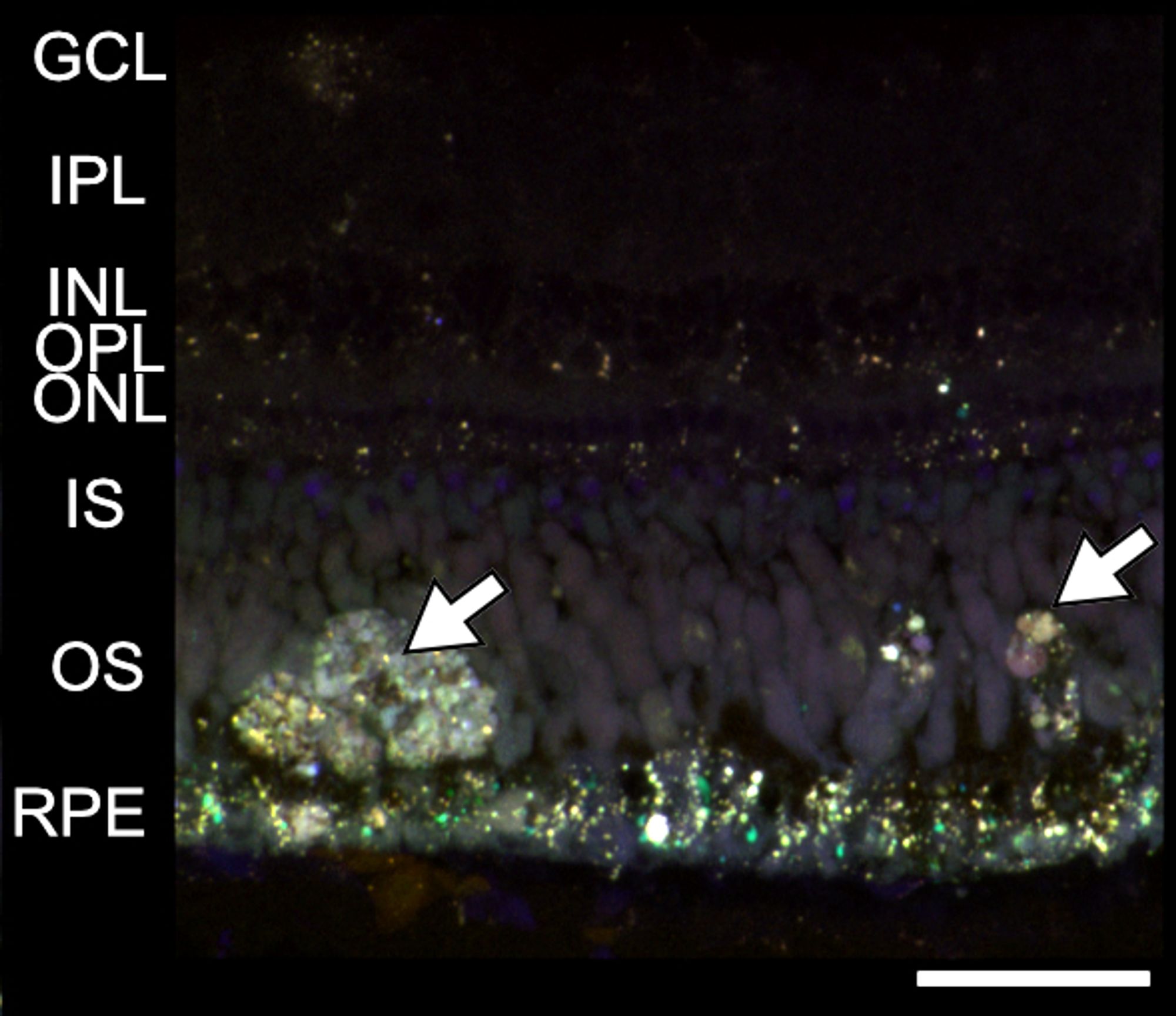
It's live! journals.biologists.com/jcs/article/... I am really proud of this work. In short, frogs develop retinal deposits that, in humans, are associated with age-related macular degeneration (AMD). This is the first step in establishing a new animal model for AMD and new therapies. 🧪 🐸

Large autofluorescent deposits (yellow, white arrows) in the outer segment/subretinal layer. In humans, deposits such as this are associated with advanced age-related macular degeneration and progression to blindness. Scale bar = 50 um.
2
DBdrbjcarr.bsky.social

I need a reading sabbatical where all I do is avoid everyone and read papers. Is that a thing? Can I make this a thing?!

0
DBdrbjcarr.bsky.social

Now this is just the cutest effing thing I've ever seen. It looks like a fat korok.

1
DBdrbjcarr.bsky.social

Mountain therapy.

A light blue stream running through a canyon. There  are rocks and tall trees on either side.
A cow elk munching on a bush with silver-grey leaves.
A lit up bridge with yellow and green trees and a mountain range in the background.
A blue waterfall surrounded by mossy rocks and green trees.
0
Reposted by Dr. Brittany J. Carr
Bbwjones.bsky.social

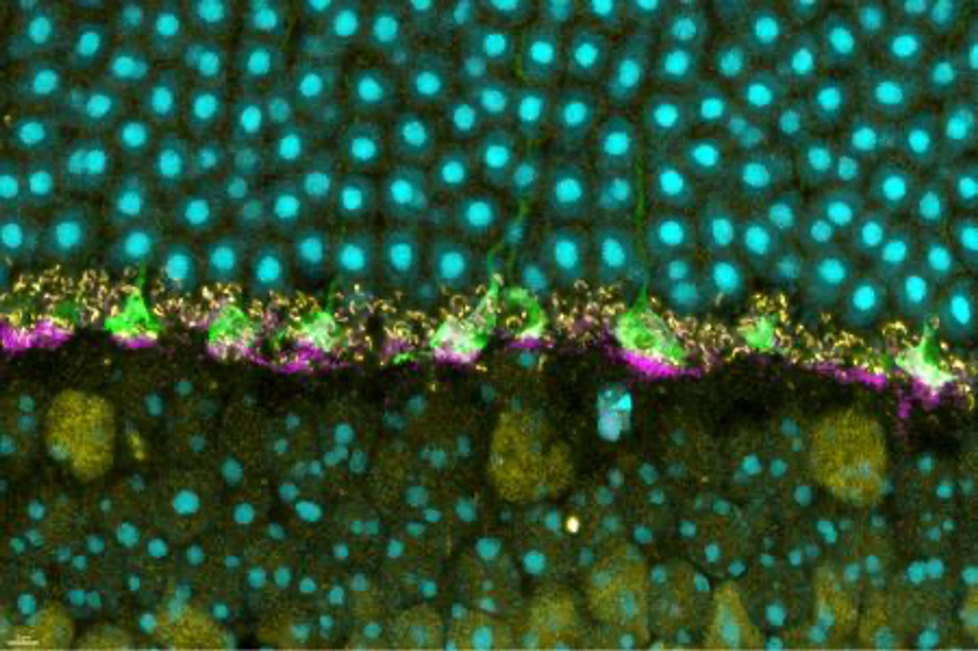
Another study that points the way towards using retina as a model of CNS neurodegeneration like Alzheimer's. Nice work vision science peeps! www.niaid.nih.gov/news-events/...

the outer layer of a mouse retina. Normal prion protein (magenta) is visible at the base of photoreceptor terminals, called cone pedicles (green). Specialized synapses are shown in yellow. The nuclei of photoreceptors are in blue above the synapses, and in blue below the synapses are bipolar cells. The cone pedicles relay information about detected light to other retinal nerve cells, which carry the information to the brain where images are constructed. The new study found that prion protein is necessary for the normal operation of the retinal synapses. Credit: NIAID
1
DBdrbjcarr.bsky.social

Lovely work. The artist did an exceptional job turning your work into art.

0
DBdrbjcarr.bsky.social

This is your arm?! Amazing artwork!

1
DBdrbjcarr.bsky.social

Hurray!

0
Profile banner
DB
Dr. Brittany J. Carr
@drbjcarr.bsky.social
Assistant Prof @UAlberta. Genetically-modified frogs 🐸, retinal degeneration, imaging. She/Her. Views are my own. 🇨🇦
372 followers452 following513 posts