Toddler with a sacral dimple Sagittal T1 MRI without contrast of the lumbar spine (left) shows hyperintense signal in the conus medullaris which lies low in position at the level of L2-L3. #FOAMed#MedEd#FOAMPed#FOAMRad#PedsRad#RadEd#RadRes#radiology#NeuroRad#PediNeuroRad#NeuroRadiology

which has associated edema in the right temporal lobe and which is causing midline shift to the left The diagnosis was subpial hemorrhage Learn more: pediatricimaging.org/diseases/sub...#FOAMed#MedEd#FOAMPed#FOAMRad#PedsRad#RadEd#RadRes#radiology#NeuroRad#PediNeuroRad#NeuroRadiology
The diagnosis was pleomorphic adenoma. Learn more: pediatricimaging.org/diseases/ple...#FOAMed#MedEd#FOAMPed#FOAMRad#PedsRad#RadEd#RadRes#radiology#NeuroRad#PediNeuroRad#NeuroRadiology#Neurology#Neurosurgery
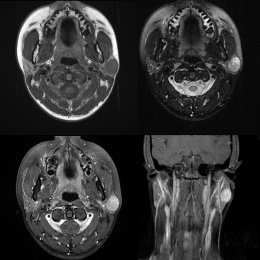
Pediatric pleomorphic adenoma radiology discussion including radiology cases.
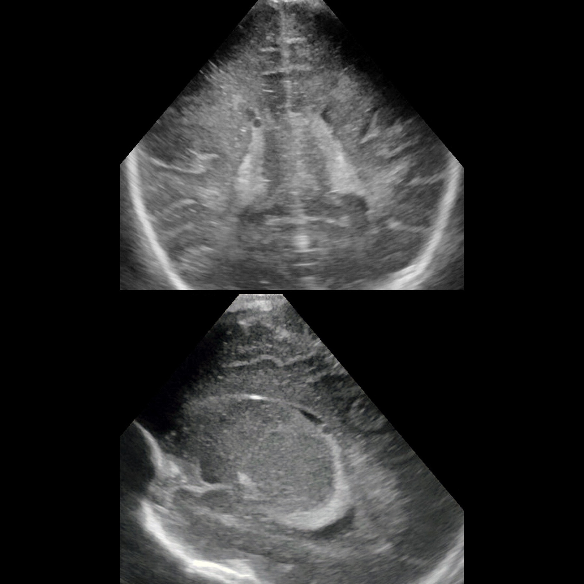
Newborn with a seizure Coronal (above) and right of midline sagittal (below) US of the brain shows a small, round, anechoic structure in the right choroid plexus. #FOAMed#MedEd#FOAMPed#FOAMRad#PedsRad#RadEd#RadRes#radiology#NeuroRad#PediNeuroRad#NeuroRadiology#Neurology#Neurosurgery
Axial T2 MRI(below) shows lesion to be in midline. The diagnosis was atretic cephpalocele. Learn more: pediatricimaging.org/diseases/atr...#NeuroRad#PediNeuroRad#NeuroRadiology#Neurology#Neurosurgery
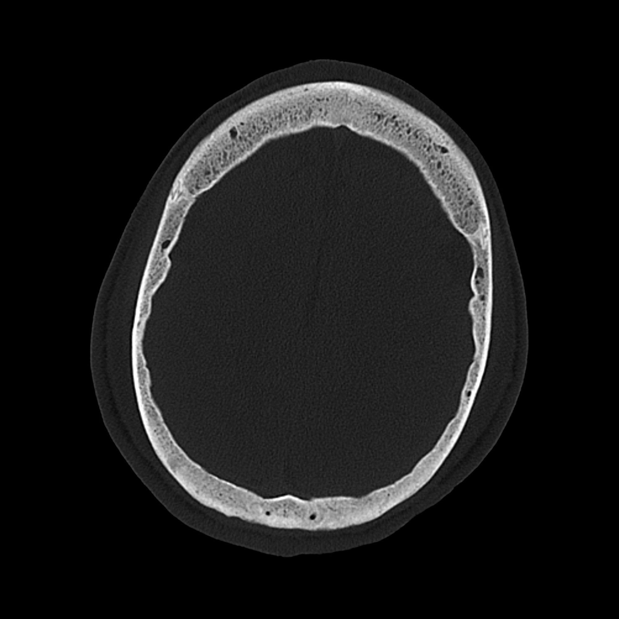
Teen with seizure disorder on anti-seizure medication CT shows diffuse thickening of diploic spaces in skull. The diagnosis was calvarial thickening due to phenytoin usage. Learn more: pediatricimaging.org/diseases/cal...#FOAMed#MedEd#FOAMPed#FOAMRad#PedsRad#RadEd#NeuroRad#PediNeuroRad
The diagnosis was Blake pouch cyst. Learn more: pediatricimaging.org/diseases/bla...#NeuroRad#PediNeuroRad#NeuroRadiology#Neurology#Neurosurgery
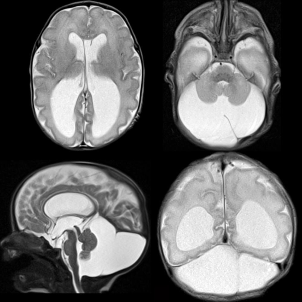
Pediatric Blake pouch cyst radiology discussion including radiology cases.
You can also follow us on social media as @pedsimaging on Bluesky(bsky.app/profile/peds...www.facebook.com/pedsimaging/www.instagram.com/pedsimaging/www.threads.net/@pedsimagingwww.twitter.com/pedsimaging/#NeuroRad#PediNeuroRad#NeuroRadiology
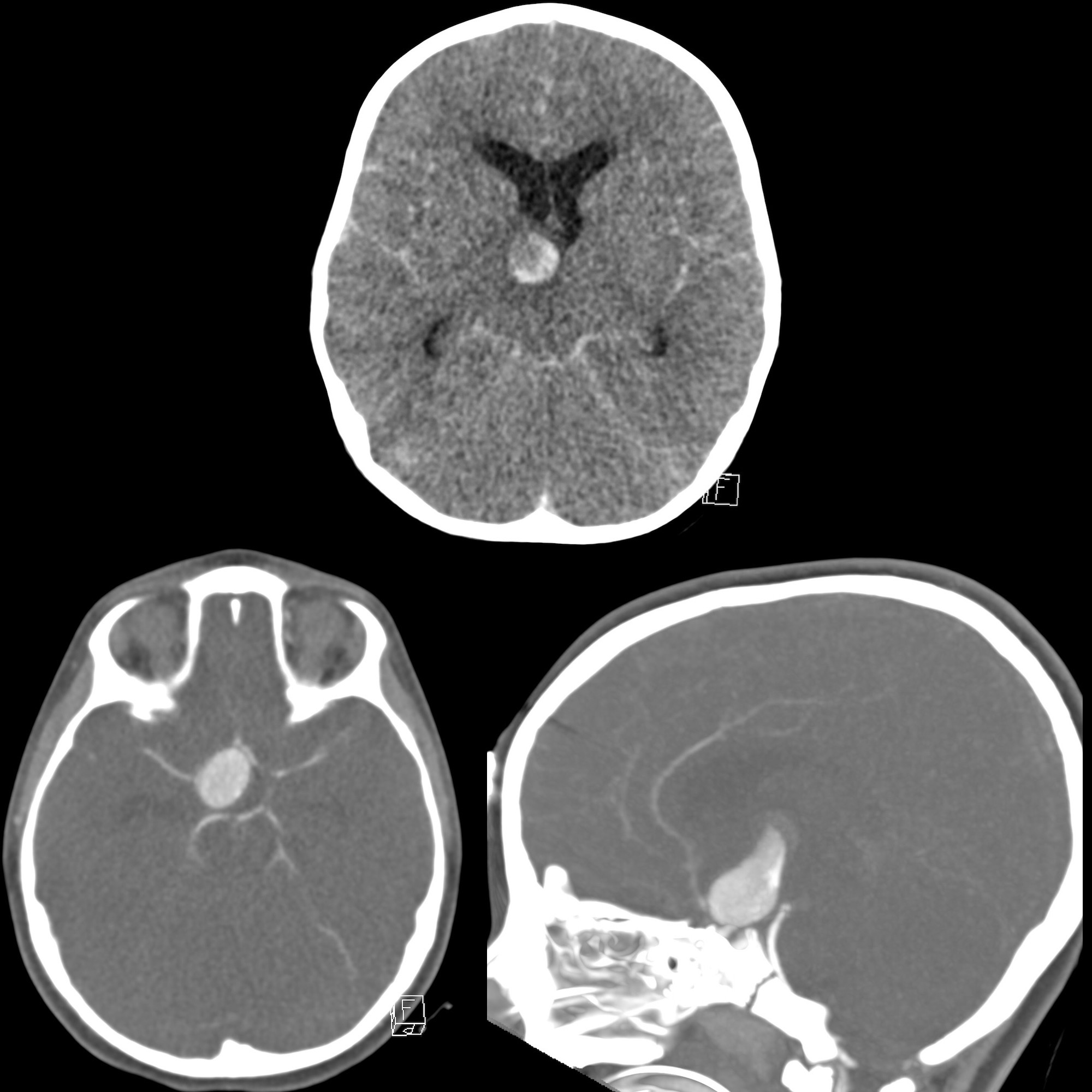
Preschooler after cardiac arrest CT without contrast(above) shows round mixed density lesion just to right of third ventricle along with diffuse subarachnoid hemorrhage+cerebral edema. #FOAMed#MedEd#FOAMPed#FOAMRad#PedsRad#RadEd#RadRes#radiology#NeuroRad#PediNeuroRad#NeuroRadiology